Meet the team
If you're interested in joining the LMS

Understanding how chemicals in our environment cause disease has been constrained by a fundamental lack of tools. We are building spatial single-cell genomics technologies and high-throughput screening platforms to reveal, cell by cell, how exposures remodel tissues and drive disease.

Every day we encounter thousands of chemicals in the air, food, and products we use. Some can damage our health by altering how our cells behave, contributing to cancer, fertility problems, and other diseases. But for most chemicals, we don’t yet know exactly how or why this happens inside the body. Our group develops technologies to read the molecular state of individual cells within their precise location in tissue, building detailed maps of how chemical exposures alter us from the inside out. Ultimately, we want to identify which chemicals are harmful and why, in order to help remove and replace them before they cause widespread disease.
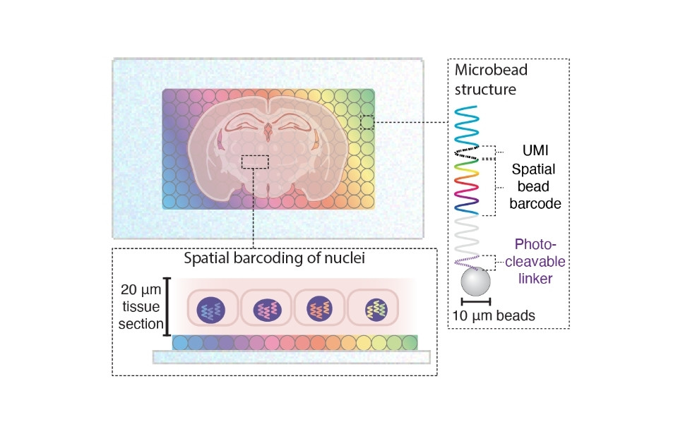
We develop and apply spatial single-cell multiomic technologies to characterise the cellular mechanisms by which environmental exposures (e.g. carcinogens, endocrine disruptors, and PFAS) perturb tissue homeostasis. Using Slide-tags, a spatial barcoding platform, we simultaneously profile somatic mutations, epigenetic state, and the transcriptome in single nuclei at spatial resolution in tissues with diverse exposure profiles and distinct disease outcomes (e.g. liver and reproductive tissues). We complement this with high-throughput in vitro screening platforms using oligonucleotide-barcoded exposures, and are exploring organoid models as a scalable alternative to in vivo experiments. Computational frameworks integrate these multimodal datasets to model exposure-driven tissue evolution, identify vulnerable cell states, and predict the effects of unseen exposures.
Environmental exposures contribute to nearly a quarter of all global deaths, yet health effects remain uncharacterised for the majority of the >350,000 chemicals in current use. By mapping the cellular consequences of specific chemical exposures at high resolution, our work will generate evidence to inform regulation and removal of harmful compounds before they cause widespread disease, including cancers, reproductive disorders, metabolic disease, and neurodevelopmental conditions. Our datasets and predictive models will support novel diagnostics identifying early cellular signatures of tissue damage. By linking chemical structure to biological effect, we also aim to inform the rational design of safer alternatives and therapeutic interventions.
Slide-tags enables single-nucleus barcoding for multimodal spatial genomics. Russell AJC, Weir JA, Nadaf NM, Shabet M, Kumar V, Kambhampati S, Raichur R, Marrero GJ, Liu S, Balderrama KS, Vanderburg CR, Shanmugam V, Tian L, Iorgulescu JB, Yoon CH, Wu CJ, Macosko EZ, Chen F. Nature·2024·:101-109·DOI: 10.1038/s41586-023-06837-4·PubMed
Spatial multiomic landscape of the human placenta at molecular resolution. Ounadjela JR, Zhang K, Kobayashi-Kirschvink KJ, Jin K, J C Russell A, Lackner AI, Callahan C, Viggiani F, Dey KK, Jagadeesh K, Maxian T, Prandstetter AM, Nadaf N, Gong Q, Raichur R, Zvezdov ML, Hui M, Simpson M, Liu X, Min W, Knöfler M, Chen F, Haider S, Shu J. Nature medicine·2024·:3495-3508·DOI: 10.1038/s41591-024-03073-9·PubMed
Regulators of male and female sexual development are critical for the transmission of a malaria parasite. Russell AJC, Sanderson T, Bushell E, Talman AM, Anar B, Girling G, Hunziker M, Kent RS, Martin JS, Metcalf T, Montandon R, Pandey V, Pardo M, Roberts AB, Sayers C, Schwach F, Choudhary JS, Rayner JC, Voet T, Modrzynska KK, Waters AP, Lawniczak MKN, Billker O. Cell host & microbe·2023·:305-319.e10·DOI: 10.1016/j.chom.2022.12.011·PubMed
The Malaria Cell Atlas: Single parasite transcriptomes across the complete Plasmodium life cycle. Howick VM, Russell AJC, Andrews T, Heaton H, Reid AJ, Natarajan K, Butungi H, Metcalf T, Verzier LH, Rayner JC, Berriman M, Herren JK, Billker O, Hemberg M, Talman AM, Lawniczak MKN. Science·2019·:eaaw2619·DOI: 10.1126/science.aaw2619·PubMed