Creating molecular technologies
We create molecular tools and technologies to address specific, intriguing challenges at the intersection of protein engineering and synthetic biology. We currently focus on two main directions:
— Protein design for therapeutics and crop protection,
— Self-sustained luminescence as a reporter tool for agricultural and biomedical applications.
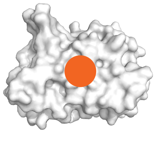
What does AI-based protein design enable in therapeutics and agriculture?
Evolutionary pressures equipped organisms with an amazing diversity of protein-based defence and attack mechanisms. A number of these have been adopted as therapeutic or crop protection molecules. Developments in de novo protein designs promise great expansion of arsenal of such proteins. We focus on solving the downstream bottlenecks in protein design pipelines and creating a robust platform for their development.
Can we use self-sustained luminescence to visualise molecular events at the organism scale?
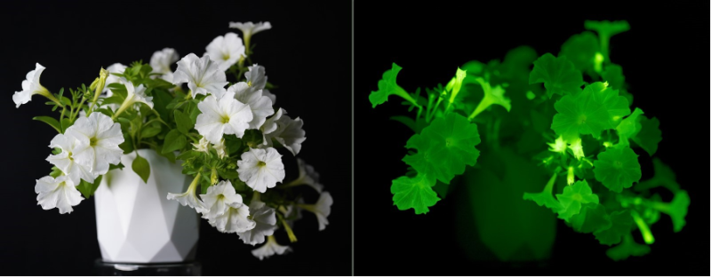
Creating organisms that glow in the dark requires understanding of biochemical pathways that lead to bioluminescence. In 2018, our collaborators and we discovered a mechanism of light emission in fungi, where bioluminescence results from activity of “caffeic acid cycle” – a short metabolic pathway catalysed by four enzymes. We showed that integration of that pathway into host metabolism allows engineering of organisms with self-sustained luminescence.
We are now working on applying self-sustained luminescence to visualise invisible physiological events. By making expression of bioluminescence genes dependent on physiological or environmental events, we can monitor virtually any molecular event completely non-invasively. For example, luminescence can show hormone activity in tissues, patterns of gene expression, or be used to create sentinel organisms that report otherwise invisible environmental cues.
For more information about the lab, visit our website: https://designing.bio