


By Sophie Arthur
August 19, 2020
Time to read: 5 minutes
A network of muscle described by Renaissance artist Leonardo da Vinci half a century ago, has been found to be crucial in our understanding of how the heart works.
An international team of researchers, led by the Computational Cardiac Imaging group at the LMS, used artificial intelligence (AI) to learn more about this 500-year-old mystery. They have shown that a complex mesh of muscle fibres that line the inner surface of the heart play a vital role in increasing the efficiency of blood flow through the heart.
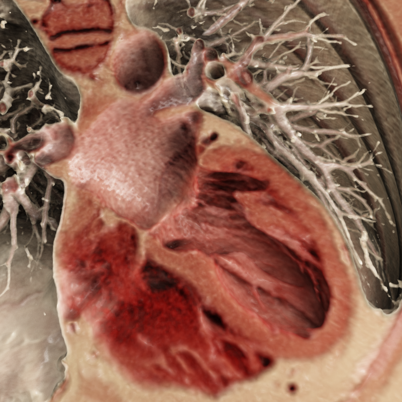
The study, published today in the journal Nature, used AI to analyse 25,000 MRI scans of the heart from the UK Biobank study. These scans revealed that unlike the smooth lining of our blood vessels and outer surface of the heart, the inside of the heart is rough, and intricately structured. This lattice lining of muscle fibres are known as trabeculae.
Trabeculae form a repeating geometric pattern known as a fractal – a repeating branching pattern that is often seen in structures across nature, from our brain cells to trees to snowflakes. One of the earliest descriptions of these branching patterns came from da Vinci in the 16th century. He was interested in why these mathematical concepts are so often seen in nature, leading to what became known as ‘Leonardo’s Rule for Branches’. He was the first to sketch these muscles in his notebooks, speculating at the time that they warm the blood as it flows through the heart. However, their true importance to human health has not been recognised until now, with the help of 21st century technology.
Today’s findings show that trabeculae are critical for the performance of the heart. They create a rough layer inside the heart which traps a thin layer of static blood along the organ’s wall. This layer reduces friction between the heart muscle and blood, and means that blood inside the heart flows more efficiently – much as the dimples in a golf ball trap tiny pockets of air, which help it to glide through the air and travel further.
Dr Declan O’Regan, Head of the Computational Cardiac Imaging group and leader of the study, said:
“Leonardo da Vinci sketched and speculated about these intricate muscles inside the heart half a millennium ago, and it is only now that we are beginning to crack this 500-year-old mystery and understand how important they are to human health.
Da Vinci was also intrigued by the link between maths and nature, so it’s fitting that we found that fractal patterns in the heart are so important for its function. This work offers an exciting new direction for our research and for understanding the role of trabeculae in disease and shows the potential for bringing together ideas in maths and biology to medical research.”
The study also discovered six regions in our DNA that affect how the fractal patterns in these muscle fibres develop. Intriguingly, the researchers found that two of these genes also regulate branching of nerve cells and whether they form straight or branched filaments, suggesting a similar mechanism at work in the developing brain as well as the heart.
The researchers found that trabeculae may influence the risk of heart disease. Using genetics to analyse data from 50,000 patients, they found that different fractal patterns in these muscles affected the risk of developing heart failure. It is hoped that this will enable future research on the disease, which affects around 920,000 people in the UK [BHF Statistics Factsheet 2020].
Dr O’Regan said:
“This network of muscles lies between fast-flowing blood inside the heart and the contracting heart muscle. The next steps for our research are to understand how these fibres affect the ‘aerodynamics’ of blood flow in the heart and how this might inform research into new treatments for heart disease. We also found that these fibres influence how fast electrical impulses travel through the heart – so they may be important for more than one aspect of how the heart works.”
Trabeculae are not static. Previous work has shown that they can change during pregnancy or with exercise, for example, so we are not born with a particular branching pattern and stuck with it. At the moment it is too early to say how different treatment regimes may affect the branching structures. But this new knowledge shows that increased understanding could have clinical applications in the future. It will be some time before this work changes the way doctors treat their patients. However, this study opens up the possibility that one day doctors will be able to improve the efficiency of the heart with drug and lifestyle changes that stem from this new understanding of trabeculae and heart failure.
Dr O’Regan discussed these potential implications:
“
It brings an exciting new avenue for my group’s research too. We want to more fully understand the interaction between the trabeculae and the flowing blood through the heart, how that contributes to the efficiency of cardiac performance and how to balance lowering the risk of heart failure against increasing blood pressure or increased risk of blood clots. That sweet spot for any future potential development of treatments needs to be found.”
‘Genetic and functional insights into the fractal structure of the heart’ was published in Nature on 19 August. Read the full article here.
Image caption: “Cardiac scan showing the mesh of muscle fibres inside the heart (Siemens Healthineers Cinematic Rendering)”.