


By Helen Figueira
May 29, 2015
Time to read: 3 minutes
Deborah Oakley
Madeline Lancaster (@MinibrainLab), who developed pioneering “mini-brains”, visited the MRC’s Clinical Sciences Centre (CSC) in West London this week, to tell CSC scientists about her latest work.
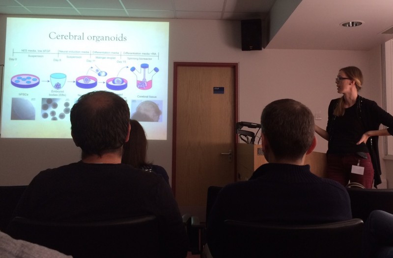
She described her two-year old technique, which coaxes human stem cells to assemble into balls of brain-like tissue. These mini-brains, known as “organoids”, contain structures that resemble parts of the brains of human fetuses up to around the ninth week of pregnancy.
“The technique has been a breakthrough,” said Simona Parrinello, who leads the Cell Interactions and Cancer research group at the CSC, and who organised Lancaster’s talk. “It’s a system where we can start to study the mechanisms and complex biology of the human brain – which we can’t do any other way.”
“The technique could be exciting for many researchers at the CSC,” said. Parrinello. “I would like to use it to recreate areas of the brain where stem cells are found.”
Lancaster’s latest work confirms that organoids could be used to study a variety of neurological diseases. In research that has yet to be published, she has shown that the nerve cells which make up organoids develop into specific brain tissues in the same sequence as nerve cells within the human brain.
“The different layers [of cells] develop in succession,” said Lancaster. “This is a hallmark of human brain development and has never been seen before.”
Her recent work also confirms that organoids contain a diverse mix of different types of nerve cells, just like human brains. “The organoids are a human system, so you can really look at human specific questions,” Lancaster said.
Scientists previously used mice as a model to study the human brain, but mice brains lack the structural complexity and intellectual capacity of our own. This has made it difficult to study human conditions such as microcephaly, which stunts brain growth and causes cognitive impairment.
“Mouse brains are only slightly affected by microcephaly,” said Lancaster. She recently used organoids to show that the condition occurs when stem cells in the brain adopt specific roles too early in development, which means that too few are left to fuel brain growth.
Lancaster, who is based at the MRC’s Laboratory of Molecular Biology, now plans to study autism and macrocephaly, in which patients’ head circumference is larger than normal. Meanwhile other teams are using organoids to study neurological conditions such as schizophrenia, and developing others types of organoids to resemble the eye, liver and colon tissue. She said, “People are generating organoids for almost any organ out there, and I think it we’ll see many more in the future.”
Take a look at a colourful organoid on our Biomedical Picture of the Day, and find out more about how to grow organoids.
—————————————————————
For further information, contact:
Deborah Oakley
Science Communications Officer
MRC Clinical Sciences Centre
Du Cane Road
London W12 0NN
T: 0208 383 3791
M: 07711 016942
E: