


By Helen Figueira
May 18, 2009
Time to read: 4 minutes
 What shocks cells into senescence?
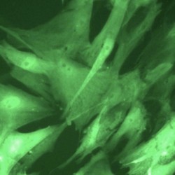
Cellular senescence – the process by which cells can enter suspended animation and stop dividing – has previously been shown to be a barrier to the spread of cancer in pre-malignant tumours.
What shocks cells into senescence?
Cellular senescence – the process by which cells can enter suspended animation and stop dividing – has previously been shown to be a barrier to the spread of cancer in pre-malignant tumours.
Senescence was first observed over 40 years ago as a mechanism that stops cells from dividing more than a finite number of times – usually about 50. It marks the onset of cellular degradation and is often seen as a cellular parallel to ageing in organisms.
In addition to this in-built replicative redundancy, cells can also be shocked into senescence prematurely by stresses, such as an oncogenic ‘insult’ sent out by a cancerous cell. This at least temporarily slows the multiplication of cancerous cells, so the study of senescence could potentially have far-reaching implications for the treatment of cancer.
Workers in the CSC’s Cell Proliferation Group, headed by Jesús Gil, have previously identified a gene called CXCR2 which mediates senescence. By inhibiting the gene, they have shown that cells can be made to live for longer. Now, they have identified the mechanism by which a locus called INK4a/Arf is derepressed by oncogenic signals to push the cells towards a senescent state.
Previous studies by other workers has shown that when the function of INK4a – normally silenced by Polycomb Group (PcG) proteins – is induced, the cells enter a senescent state prematurely.
In order to elucidate the mechanism by which this occurs, the researchers developed an ‘inducible’ system that allowed them to introduce oncogenic signals to human and animal cell cultures in vitro. Cells receiving an ‘insult’ developed characteristics of senescence, followed by a reduction in the methylation levels of a particular amino acid – ‘lysine 27’ on histone H3 – which must be methylated for the PcG proteins to bind and repress INK4a. Demethlyation of the amino acid ultimately leads to derepression of INK4a in human breast fibroblast cells, eventually leading to cellular arrest and the transmission of the ‘feedback’ senescence signals to stop growth. A slightly different pathway in mouse embryonic cells, where both INk4a and Arf are activated by the insult, echoed the result in humans, but showed that there are important differences between human ARF and its counterpart in mice.
The team reasoned that, besides nucleosome exchange, the reduction in methylation levels of the Ink4a/Arf locus could result either from a reduction in the levels of the methyltransferase enzyme EZH2 – which adds methyl groups to lysine 27 and normally facilitates PcG binding – or by an increase in either UTX or JMJD3 demethylase, which remove methyl groups and is important for cell differentiation. They followed the levels of these proteins by immunofluorescence, and determined that levels of JMJD3 increased over a period of days, whereas EZH2 increased. UTX level did not change, ruling out the involvement of the protein in the senescence pathway.
The suggestion that JMJD3 may be an important tumour suppressor seems to be backed up by clinical evidence: Elevated levels of the protein are often observed in benign and premalignant lesions, whereas aggressive tumours show a decrease in JMJD3, characteristic of a cancer in which the safeguarding measures of senescence have been overcome by the ravages of cancer growth.
Stefan Janusz
This work was published in Genes and Development.
Barradas, M., Anderton, E., Acosta, J.C., Li, S., Banito, A., Rodriguez-Niedenfuhr, M., Maertens, G., Banck, M., Zhou, M.-M., Walsh, M. J., Peters, G., and Gil, J. (2009). Histone demethylase JMJD3 contributes to epigenetic control of INK4a/ARF by oncogenic RAS.Genes Dev. 23 1177-1182.
Also see: Agger, K., Cloos, P. A., Rudkjær, L., Williams, K., Andersen, G., Christensen, J., Helin, K., May 2009. The h3k27me3 demethylase jmjd3 contributes to the activation of the ink4a–arf locus in response to oncogene- and stress-induced senescence. Genes & Development 23 (10), 1171–1176. (Same issue as above).