Scientists at the Laboratory of Medical Sciences and Imperial College London have discovered a missing link in how vegetables benefit our health: chemicals from green vegetables like broccoli can help maintain the normal function of blood vessels in the small intestine, which in turn helps protect us from infection. This study was published today in Nature.
The blood vessels are the transport network of the body, ferrying oxygen and nutrients to our organs, and transporting immune cells to where they are needed most. Keeping these blood vessels functioning normally is imperative in keeping all our organs in good health, but surprisingly little work has gone into identifying what these maintenance signals are.
“As a lab, we are interested in the consequences of nutrient sensing in health and in inflammatory disease”
said Dr. Chris Schiering who led the study.
“We asked whether there is a connection between the diet and the normal functioning of blood vessels in the gut”.
At the heart of this communication between the food we eat and our cells, is a molecular sensor called the ‘aryl hydrocarbon receptor’, or AHR for short. The AHR sits inside our cells and works by sensing chemicals derived from vegetables like broccoli, cabbage and kale. Sensing these chemicals leads to changes in the genes the cell makes that in turn changes the cells’ behaviour.
“Studying AHR is exciting as it forms a direct molecular link between the foods we eat and reprogramming the cells in our gut”
first author Dr. Ben Wiggins explained.
“What we found was quite surprising – the cells that make up our blood vessels use AHR to sense these vegetable-derived chemicals, and it is exactly this process that helps maintain the blood vessels normal function, and consequently, a healthy gut!”.
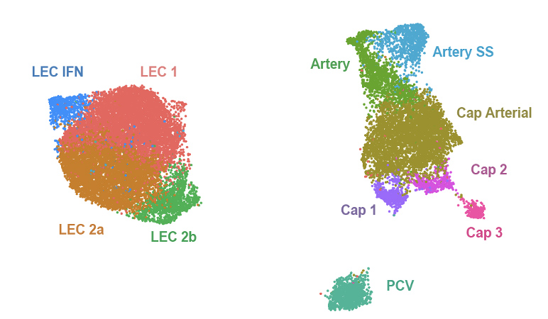
The work started with comprehensive profiling of all the cells that make up the blood vessels – so called ‘endothelial cells’. By sequencing the genes of thousands of individual endothelial cells from mice, the team uncovered the complex variation in these cells within the gut, finding new subtypes that contribute to normal vessel function in subtly different ways (Fig.1).
Fig.1 – ‘the Turtle and the Duck’ shows the groups of different endothelial cell subtypes uncovered by the gut. Each dot shows one cell and different colours show the different subtypes along with their names. The closer two subtypes are together on the plot, the more similar the genes they produce.
“Another benefit of our work is the detailed description of new endothelial cell subtypes that have never been seen before” Chris said. “This will ultimately help other scientists understand the gut blood vessels like never before”.
Next, the researchers showed that when the blood vessel cells used AHR to sense the dietary chemicals, AHR turned off genes involved in both cell division and inflammation. The study demonstrates this process happens in both mice, and human cells. This is important to keep the vessels in a resting and calm state, to prevent the relentless inflammation that is part of many chronic diseases.
So, AHR in blood vessels sense vegetable-derived chemicals, and this process is used to keep those vessels healthy and under control.
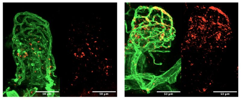
But what happens when this goes wrong? The team addressed this by generating a mouse model where AHR was deleted in endothelial cells. Using these mice, they showed that without AHR to keep your vessels in check, they now undergo excessive cell division (Fig.2), excessive inflammation, and even were more susceptible to gut infections. This highlights the importance of keeping the blood vessels in check for the health of the whole organ.
Fig.2 shows the blood vessel network (green) of genetically normal mice (left images) compared to those lacking AHR in their blood vessels (right image). Mice without AHR in their blood vessels had far more blood vessel cell division signals, shown as the red staining.
When asked exactly how this might work in infection, Ben explained “by turning off these unnecessary inflammatory genes, the blood vessels can alter the types of immune cells recruited into the infected organ. This is important as the wrong type(s) or the wrong number of immune cells that enter our organ can cause unwanted damage”.
“It is all about balance when it comes to inflammation. Too little and the infection will not be dealt with, but too much and the damage caused by the immune system can do more harm than good. Our study suggests AHR works in blood vessels to fine tune this level, allowing appropriate and optimum responses to infection.”
– Dr Benjamin Wiggins, first author of the publication.
This theory is supported by work published in tandem that also investigated AHR in blood vessels, but this time within the lung.
Their study, led by Jack Major and Andreas Wack at the Francis Crick Institute, demonstrated that AHR in lung endothelial cells was required for maintaining a healthy barrier within our lungs and in aiding appropriate immune responses after lung infection. Read more about AHR’s role in lung endothelial cells.
This work was done by MRC LMS in collaboration with Imperial College London. The research was published in Nature and funded by the Medical Research Council (MRC), Imperial College London and The Wellcome Trust.