


By Helen Figueira
May 25, 2010
Time to read: 2 minutes
 Mary Rutherford interviewed for NewScientist magazine
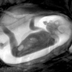
The Perinatal Imaging group’s groundbreaking work on in utero scans of babies has featured in the popular press before (see First 3D MRI Scans of Unborn Babies). Recently, a rather fun article by Linda Geddes on the NewScientist website charts her experiences with her own prenatal progeny: a “juvenile Jackie Chan”, “boxing clever” as she takes a “run up” against the uterine wall.
The article highlights some of the important science behind the Perinatal Imaging group’s use of MRI, especially the advantages of MRI over the more conventional ultrasound in illuminating detailed foetal physiology and neurology during the later stages of pregnancy.
“With ultrasound you have a relatively limited view, and beyond 32 weeks you can only really see parts of the body moving,” says Mary Rutherford in the article. In comparison, MRI can produce high resolution images, elucidating fine structures while the baby continues her ‘work-out’ in the womb.
One interesting finding highlighted in the article is that the difference between the foetal acrobatics of a 20 week-old and a 40 week-old are minimal, suggesting that these ‘kung-fu’ moves are actually the result of “fairly primitive brain processes”.
Mary Rutherford added: “People had thought that as gestation developed, babies’ movements would become more complex as a sign of higher brain activity, but that doesn’t seem to be the case.”
Linda Geddes’ article appears on the NewScientist website here