


By Helen Figueira
November 30, 2009
Time to read: 1 minutes
 Perinatal Imaging
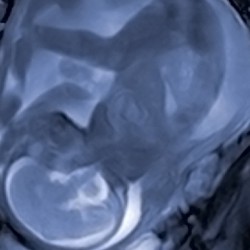
30 November 2009 Cutting-edge 3D MRI techniques being pioneered by the CSC’s Perinatal Imaging group are reported in an article on the BBC website by BBC Health Reporter Jane Elliot.
From the article:
Baby Miller makes his first appearance on screen.
He can be seen moving and swallowing – a proud moment for parents-to-be Sian and Brian, and a welcome addition to their baby memorabilia.
The 3D scan shows that the baby is coming on well, and his development is normal.
But the scan is more than just a memento.
For the new Miller is one of the latest foetuses to be enrolled in a brain study at London’s Hammersmith Hospital, in collaboration with the Medical Research Council.
…
Professor Mary Rutherford said her team had got round this by taking multiple scans of the brain and then slotting them together to make a 3D image.
“This information will help obstetricians to decide whether a baby is likely to have severe problems with development or whether to deliver a baby sooner as brain growth may be better outside the womb,” she said.
You can read more of Jane Elliot’s article and watch the video on the BBC website here.