


By Staff Member
December 14, 2022
Time to read: 4 minutes
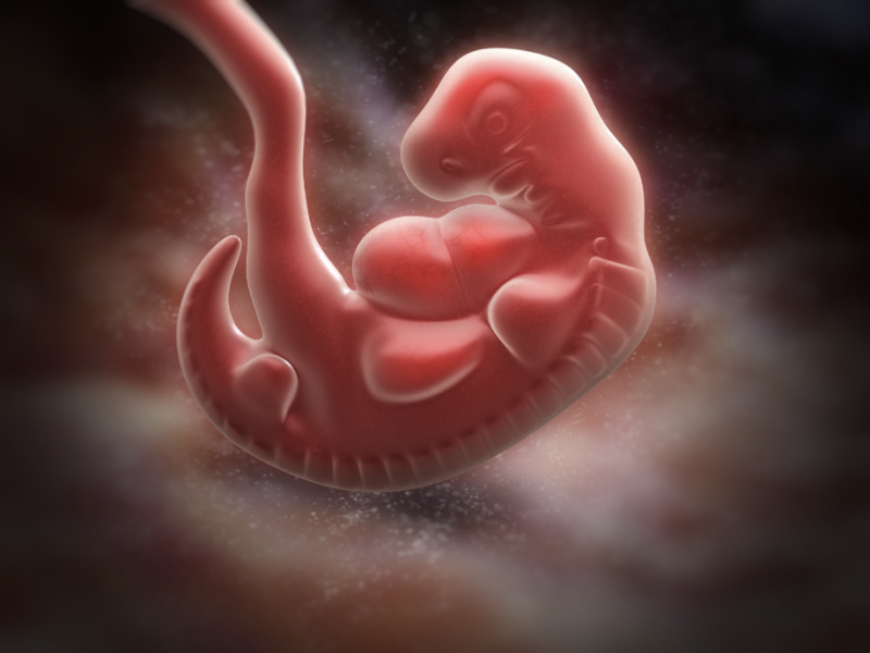
Researchers uncover molecular events that promote human spinal cord development using embryonic stem cells
Leader of the MRC LMS Development and Transcriptional Control research group Dr Vicki Metzis together with collaborators from the University of Dundee and The Francis Crick Institute uncovered how changes in a molecular signal can drive the formation of the spinal cord. Using mouse embryos and human embryonic stem cells to model human development in vitro, researchers were able to uncover the mechanisms that promote the generation of the spinal cord. This work provides vital fundamental knowledge to forward the development of regenerative medicine to repair, replace and restore damaged cells in the nervous system.
The human body is made up of at least 200 cells. This means that ‘the right cell types need to form in the right place, and of course in the right proportion’. Each cell in the embryo has a designated ‘fate’ that determines which cell type it becomes.
Dr Vicki Metzis and the research teams involved in this study are particularly intrigued by how cells in the embryo form the spinal cord. Understanding how the activation of neural genes in progenitor cells can lead to the formation of the spinal cord provides fundamental insights that may also inform regenerative medicine approaches in the future. This is particularly important as damage to the spinal cord can cause severe and long-lasting health consequences. In the team’s most recent paper they found that in the earliest phases of spinal cord development, inhibition of the ERK1/2 signalling cascade leads to a dramatic change that makes neural genes in the genome easier to ‘reach’, ‘read’ and activate.
“Our work takes an important step forward in our understanding of the molecular events that drive spinal cord development in humans. We hope that by understanding the basic principles behind building tissues like the spinal cord, we may be able to apply those insights to help reconstruct diseased or damaged tissues in the future”
– Dr Vicki Metzis, co-corresponding author of this publication
DNA is organised into chromosomes by being tightly coiled around and amongst proteins. This complex of proteins and DNA is referred to as chromatin. Therefore, certain molecular signals are needed to uncoil this DNA to reach the genes that need to be activated and ‘read’ by the transcription factor proteins. Dr Vicki Metzis’ previous work in the lab of Dr James Briscoe at The Francis Crick Institute had shown that distinct changes to chromatin accessibility are vital to initiate spinal cord development in mice. Dr Claudia Semprich, the lead author of the new publication working in the laboratory of Professor Kate Storey at The University of Dundee, identified changes occurring at the neural gene Pax6 in both mouse embryos and human spinal cord cells. This raised the intriguing possibility that similar mechanisms may control spinal cord development in these two species. Working together, the researchers tested this idea by examining how broadly these effects might be taking place throughout the genome.
The researchers used human embryonic stem cells to monitor how cells develop into spinal cord progenitors, applying sequencing techniques to identify the location of accessible chromatin. It was found that the loss of ERK1/2 activity triggered synchronous changes in chromatin accessibility at thousands of neural genes.
In their experiments, the researchers discovered that blocking ERK1/2 signalling caused neural genes to be activated prematurely. The molecular events taking place after blocking ERK1/2 signalling were not reversible, even when the signalling pathway was reactivated. This indicates that such a change in signalling provides directionality to cells to progress spinal cord development.
These findings highlight similarities between the early stages of mouse and human spinal cord development and raise important questions about how these events are finely controlled in the genome. Discoveries in this area have been made possible through the use of embryonic stem cells to model development, and are crucial to advance human biology, disease modelling and regenerative medicine.
During this research, Dr Vicki Metzis went on to lead and set up her own lab at the MRC LMS. She and her team, The Metzis Lab, continue to investigate the intricate molecular mechanisms that determine a cell’s fate with the ambition to contribute valuable insights for regenerative medicine, using the latest advances in embryonic stem cells, genome engineering and mouse genetics.
This paper was published by PloS Biology and was supported by funds from the Biotechnology and Biological Sciences Research Council, the Francis Crick Institute, the Wellcome Trust and The Royal Society.
This research was performed in collaboration with The University of Dundee, The Francis Crick Institute and Imperial College London.
This news article was written with Sofia Velazquez Pimentel