By Helen Figueira
July 26, 2012
Time to read: 4 minutes
 Elucidating the molecular mechanism for DNA repair
Scientists estimate that every cell in our body is subject to as many as a million molecular lesions a day, simply due to the daily rigor of life and exposure to the environment. Lesions that target the DNA are of particular concern. Contained within the twists and turns of the DNA helix is the instruction manual for life at the cellular level. Breaking this helix spells disaster for the cell in which it is housed, leading to mutations, disease and sometimes death. Consequently cells have evolved mechanisms to patch up damaged DNA. When a break occurs that cuts through both strands of the helix, the cell attempts to seal the gap by sticking a similar piece of DNA back in, through a process called recombination. New research from the Cell Cycle group at the CSC, published in Current Biology, reveals how the protein complex, cohesin, lends a helping hand.
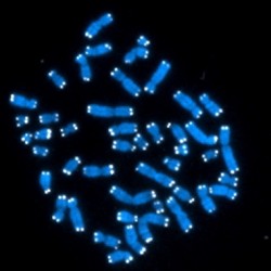
Before a cell divides it duplicates its chromosomes, with each half termed a sister chromatid. Cohesin physically tethers these chromatids together until it is time for them to separate during cell division. This ensures the correct segregation of genetic information when the chromosomes finally pull apart and populate the newly divided cells. Should this process fail, a state of aneuploidy can ensue, where the chromosomes become unevenly distributed in cells, often leading to disease.
The chromosome-binding exploits of cohesin are not only vital for the faithful segregation of DNA during cell division, but also for a special kind of DNA repair called sister chromatid recombination. “When you have a DNA double-strand break in a chromatid, the most efficient way to repair it is by using its sister chromatid because it contains the same genetic information. Ring-shaped cohesin facilitates this by tethering the chromatids together”, explains CSC’s Luis Aragon, who uses the model system of yeast to investigate this process.
Aragon’s findings reveal how SUMOylation enables cohesin to fulfil its role in DNA repair. “When a double-strand break occurs, cohesin becomes SUMOylated. When we prevent its SUMOylation, repair is blocked.”
Ready, set, SUMOylate
SUMOylation is the modification of a protein, after it has been translated, through the attachment of small proteins called SUMO (Small Ubiquitin-like Modifiers). The enzymatic cascade leading to SUMOylation begins with proteases that prime SUMO for binding and is followed by a series of enzymes named E1, E2 and E3 that are responsible for the activation, conjugation and ligation of SUMO, respectively. The final stage of ligation results in the attachment of SUMO to its target protein. SUMOylation regulates a range of critical cellular processes, including, cell death, transcription and the cell cycle. Consequently defective SUMOylation has been implicated in a range of diseases including neurodegeneration, cancer and diabetes.
SUMOylation is a post-translational protein modification that requires several enzymatic steps, one of which is mediated by the SUMO E3 ligase family of enzymes. The protein complex, Smc5-Smc6, which contains one such enzyme, is responsible for the SUMOylation of cohesin. When cohesin was mutated so it could no longer be SUMOylated, it was unable to facilitate double-strand break repair. “Cells containing mutant cohesin are therefore more sensitive to DNA-damaging agents,” says Aragon.
Co-operation between Smc5-Smc6 and the cohesin complex is fundamental to the DNA damage response. The failure to repair double-strand breaks results in a variety of disease pathologies, including immunodeficiency, neurodegeneration and cancer. Maintaining the integrity of the genome through DNA repair is therefore vital to prevent the onset of genetic diseases.
-LF
Reference:
McAleenan, A., Cordon-Preciado, V., Clemente-Blanco, A., Liu, I.-C. C., Sen, N., Leonard, J., Jarmuz, A., Aragón, L., Jul. 2012. SUMOylation of the α-Kleisin subunit of cohesin is required for DNA Damage-Induced cohesion. Current biology 2012 | Abstract
Image credit:Published under Creative Commons, Hesed Padilla-Nash & Thomas Ried