


By Staff Member
May 24, 2024
Time to read: 5 minutes
Bioluminescence imaging borrows the firefly’s capacity to generate light and imbues living cells with the ability to glow. The technique has now been used to examine how external factors influence Aryl Hydrocarbon Receptor (AHR) activation, a protein that acts as a crucial environmental sensor within the body. Cyp1a1 is a gene directly dependent on the activation of AHR, and by tracking illumination linked to this gene’s activity, the team discovered prolonged AHR activation in the lungs. The approach could be used as a living sensor to evaluate air conditions and track the immediate health implications of a changing environment.
Read the article in Communications Biology: https://doi.org/10.1038/s42003-024-06089-6
 With a little genetic engineering, cells in the body can glow like a power button shining green when switched on. This bioluminescence is at the heart of a new cross-group collaborative study from the MRC Laboratory of Medical Sciences (LMS) investigating how environmental and dietary factors can influence gene expression and ultimately health. “We created a reporter mouse to detect, through bioluminescence, gene activation in response to environmental substances,” said co-author Nicolas Veland. This mouse model visibly demonstrated, through light expressed by its cells, when a particular gene was activated in response to different environmental stimuli.
The team used this setup to explore fluctuations in the genetic response to external factors, in the process revealed uniquely persistent changes that occur within the lungs, and created a system that could be used as a live monitor of environmental conditions.
Central to the research is the Aryl Hydrocarbon Receptor (AHR), a protein that regulates the cellular response to external cues. “AHR is a receptor that is normally inactive but quickly becomes activated upon exposure to chemicals from our diet, contaminants from the environment or biproducts of our microbiome,” said co-author Hannah Gleneadie. “Essentially, it is a sensor of the environment that controls the cellular response to it.”
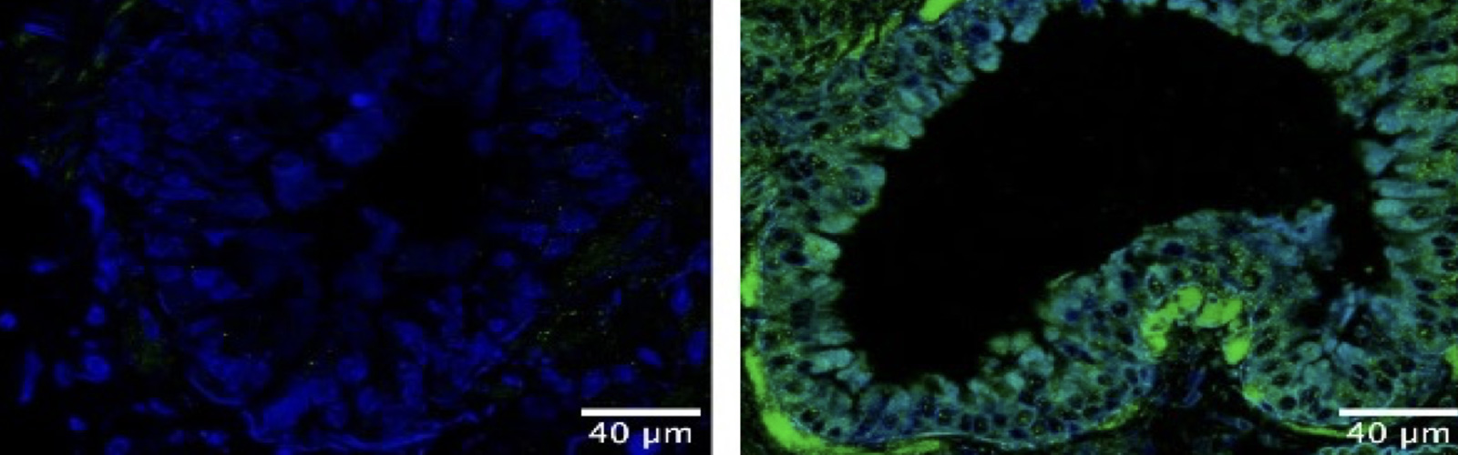
The team examined the expression of a gene, Cyp1a1, that is directly dependent on the activation of AHR, so were able to watch how AHR activity changed at different time points after exposure to chemicals. “Using this approach, we can see AHR-dependent gene activation in live animals, and can image the same mouse at different time points after exposure to AHR-activating chemicals,” said Gleneadie.
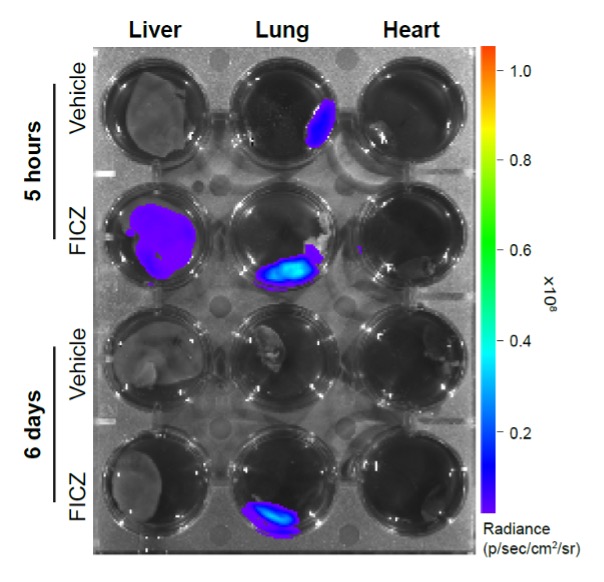
Using this approach the team saw unusual AHR activity in cell populations in the lungs. “To our surprise, we found that activation of AHR was prolonged in the lung,” said Veland. “Upon exposure to chemicals that get cleared rapidly in the body, Cyp1a1 expression was maintained in the lung but not elsewhere in the body.” AHR can be beneficial, involved in controlling disease, or detrimental if its expression gets out of hand. In this case, said Veland, “the prolonged AHR activation in the lung suggests that our lungs are very sensitive to environmental substances and this effect on the lungs could directly impact our health.”
his technique creates an instant, visual record of how environmental or dietary elements are affecting the body. “The fact that AHR is important for human disease, including cancer and autoimmunity, makes this tool very valuable as it could be applied to any context where you want to monitor AHR activation, either by testing chemicals that induce it or, when it is exacerbated, through testing inhibitors to inactive AHR,” said Veland. “All these screenings could be performed in living mice and cells. Perhaps, our reporter could be used as a living sensor to evaluate air pollution conditions in order to extend ULEZ.”
The work reveals important information about the susceptibility of the lungs to lasting harm from environmental factors, but the promise of the technique is equally significant. “We hope that this tool would be beneficial for the scientific community and would help to understand better the impact of the environment on our genes and what can we do to improve it from a human health perspective,” said Veland.
This study is the latest result of years of work from both the Epigenetic Memory Research Group and many others at the LMS, and is just one practical application of the approach. Refining and developing the tool will yield even more opportunities to illuminate the mechanisms underpinning health and disease:
“Currently, detection of bioluminescence depends on injection of the substrate, luciferin. An exciting future step in bioluminescence research is to make an autoluminescent mouse, that expresses both the enzyme and the substate required to generate light (as happens in nature in fireflies),” said Gleneadie. “This would allow real-time detection of gene expression changes, in a living, moving animal.”
While work continues on the technical approach, there remain questions about the AHR discoveries: “In terms of the AHR study, the next step in this work is to understand exactly what is causing the prolonged activation of AHR in the lung,” said Gleneadie. With an expanding toolkit and pressing questions about the mechanisms of health and disease, there will be much more to see on the way.