By Sophie Arthur
October 28, 2020
Time to read: 7 minutes
By Dr Sophie Arthur
The gut-brain axis describes the two-way communication between the neurons that make up the central nervous system, which includes the brain, and the so-called peripheral nervous system, which includes the gut. There is increasing evidence that the brain can influence gut health and function, and vice versa. Researchers are uncovering how this communication link between the gut and the brain is essential for maintaining energy balance and in regulating appetite.
For issues such as the obesity crisis, exploring how the gut-brain axis maintains energy balance could help us to understand what might be going wrong when appetite isn’t well regulated. The same is true for pregnancy and in reproduction. Across many animal species, including flies and humans, reproduction induces an increase in the amount of food a female might consume. Creating healthy offspring puts additional metabolic demands on a female’s body, so increasing food intake helps many species to cope with these needs.
A team at the MRC LMS has found that fruit flies increase their food intake during reproduction as a result of changes in the activity of the neurons that ‘talk’ to the gut. These findings help to explain how the increased food intake of mothers sustains their progeny, but when it is disrupted could lead to infertility or weight gain.
A mother’s hunger
Fruit flies have been used to study how nervous systems form and function for decades, but looking at the nervous system of the gut has received less attention. This research group from the LMS has been describing these connections between the brain and the gut in flies for several years, building a picture of how the gut-brain axis helps to control key decisions and physiology.
In their latest study, published today in the journal Nature, the Gut Signalling and Metabolism group reveal that a process called neuronal remodelling – changes in how some neurons function – is key in enabling female fruit flies to meet the increased metabolic demands of producing healthy offspring. They reveal that the gut-brain axis is involved with the regulation of food intake and provide a mechanism by which this is happening involving the neurons that ‘talk’ to the gut.
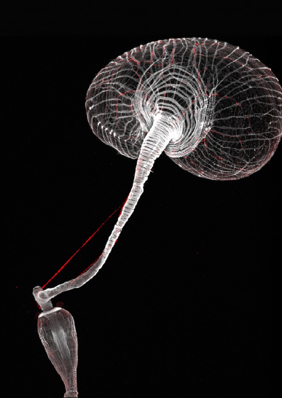
The team, along with their collaborators at NIH in the USA, ESPCI in Paris, MRC LMS and Queen Mary’s University London, has found that an expandable digestive organ in the gastrointestinal tract of fruit flies, called the “crop”, is regulated via a complex interchange of messages involving so-called enteric neurons. These neurons are known to mediate communication between the gut and the brain.
Interestingly, the crop is present only in adult fruit flies, and not in the juveniles. Little was known about this structure, but observations from the lab over the years have shown that this stomach-like structure is more than just a storage organ.
During this latest study, enteric neurons were found to trigger changes in the crop but only in female flies and only if they had “mated”. The team tested this observation by modulating the activity of these neurons in both males and females, and in both virgin and mated flies, and they found that only mated females were affected.

The enteric neurons stimulate the crop after detecting signals from two places in the mated females. The first is a steroid hormone, which probably comes from the ovary, similarly to oestrogen in humans. The second is a so-called enteroendocrine hormone made by the gut itself. Neither set of hormones were known to be ‘talking’ to the gut, and this enteroendocrine hormone was not known to be regulated by mating.
The enteric neurons contain receptors for both sets of hormones. The neurons in mated females appear to “sense” the change in the amount of hormones present after mating. When the hormones bind to the receptors on the neuron, it would fire and cause a change in the way it releases a neuropeptide – a small protein released by neurons – called myosuppresin.
The myosuppresin acts on the muscles of the stomach-like crop, relaxing these so that it can expand and allow an increase in food intake. In a collaboration with the Behavioural Genomics team at the MRC LMS, the team developed a fluid dynamics model to assess how the expansion of the crop could result in increased food intake, before testing it experimentally. Due to the negative pressure in the crop, flies draw up food via suction. The researchers found that in mated females where the crop had expanded, those flies were able to ingest more food by taking bigger sips because of the increased ’suction power’ of the enlarged crop. Like many other animals including humans, female flies were known to increase food intake during reproduction, but the neuronal mechanisms involved were unknown, and further work is required to fully understand these fluid dynamics of the crop.
The tip of a reproductive iceberg?
This latest research builds on a previous study from the group and provides further insight into how a body adapts in preparation for pregnancy. In a study published in 2015, the team described how a hormone, called juvenile hormone, is released during pregnancy and triggers the gut to physically become larger to anticipate additional metabolic demands. This meant that without an increase in food intake, the fly could extract more nutrients from the food in order to support the growth of healthy offspring.
Irene Miguel-Aliaga, Head of the Gut Signalling and Metabolism group and senior author on the study, discusses how today’s finding build on the lab’s earlier findings:
“Five years ago, we found what we now believe was the tip of an iceberg: the maternal intestine grows in size during reproduction, and this is required to sustain the developing offspring. We have now learned that the nerve cells that control this intestine also ‘know’ that the female fly is reproducing, and make the fly eat more. So, knowing that the female fly is reproducing, the intestine changes both itself and the behaviour of the fly to help the offspring develop.”
“I now wonder how many other types of cells are reproduction-sensitive, and how they communicate to sustain reproduction and/or change maternal behaviours. I also wonder whether male cells also know that the male has mated, and to what extent that affects the physiology and behaviour of males during and after reproduction. Finally, are these mechanisms relevant to animals such as humans with a placenta (an organ dedicated to reproduction)? Do our intestines also grow? Do the nerve cells on our gut also change? Is all of this reversible? One answer always leads to many more new questions.”
It remains unknown whether and how these two processes of gut growth and neuronal remodelling coordinate, and if the order in which each hormone acts is important. What is clear is that if either process is prevented, reproductive output decreases in fruit flies.
“From a fly perspective, we want to understand the fundamental links between gut resizing and neuronal remodelling. Do these two events happen independently in a female during reproduction, or does one enable the other perhaps? More generally, and for any potential future therapeutics, we want to explore whether the neurons that control the mammalian gastrointestinal tract are also sensitive to sex or reproductive cues.
Dafni Hadjieconomou, first author of this study, shared more about the findings:
“Although this is several steps down the line, and requires significant and important basic scientific research first, this knowledge could potentially lead to therapeutics that target these neurons that signal to the gut. This could help in a variety of different situations where excessive food intake may be an issue such as in the obesity crisis we see in today’s society, or in helping to explain why many women gain weight during pregnancy and struggle to lose it. But such advances will only be possible if we can understand what happens at the molecular level under normal circumstances and when dysregulated.”
‘Enteric neurons increase maternal food intake during reproduction’ was published on 28 October in Nature. Read the full article here.