


By Helen Figueira
June 28, 2012
Time to read: 5 minutes
 CSC Scientist Wins Young Investigator Award
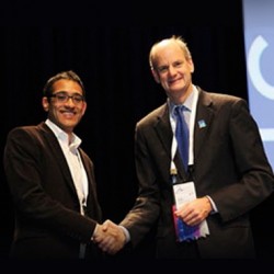
Taking a novel approach to one of the prevailing problems of magnetic resonance imaging (MRI) has allowed Shaihan Malik to peek inside the body with newfound clarity. Now his research, carried out in the Imaging Physics and Engineering group at the CSC, has earned him the prestigious 2012 I I Rabi Young Investigator Award at the 20th Annual Meeting of the ISMR
“It’s a real honour,” said Shaihan, reflecting on the achievement. “ISMRM is a large international community, and the premiere society in my field, so this means a lot.”
Shaihan’s research, carried out during an MRC Career Development Fellowship at the CSC, aimed to improve the clarity and consistency of the images produced by MRI scans. Atomic nuclei within our body, such as the hydrogen nuclei in water molecules, ‘spin’ like microscopic gyroscopes. This spinning electric charge behaves like a tiny bar magnet. When in the strong magnetic field of the MRI machine, these tiny magnets align to match the surrounding field. During MRI scanning, pulses of radio waves disrupt this alignment, and this alteration is interpreted to give the familiar black and white images of MRI scans.
This is not, however, straightforward. Your body is a sack of conductive fluid and matter, which unevenly distorts the radio waves. As a result, unpredictable bright and dark tinting often obscures the images produced.
One attempt to combat this inconsistency is a new technology called Parallel Transmission: instead of a single source of radio waves, there is an array of transmitters around the patient, which can each be independently controlled. Most attempts to harness this new technique have tried to create a uniform field around the whole body. Shaihan, however, took a different approach. Accepting that creating a totally uniform field is still beyond the capacities of the technology, Shaihan intentionally varied the field patterns. If you first ascertain how the different areas of the body will be affected by the waves, you can alter the radio wave output so that a uniform image is formed.
“Instead of trying to make uniform fields, we tried to make uniform images,” explains Shaihan. “A lot of the research in this field is very focussed on the technology itself. We tried to make it more clinically relevant, by concentrating on the image quality.” This focus on clinical application has yielded impressive results:
CSC Scientist Wins Young Investigator Award
Taking a novel approach to one of the prevailing problems of magnetic resonance imaging (MRI) has allowed Shaihan Malik to peek inside the body with newfound clarity. Now his research, carried out in the Imaging Physics and Engineering group at the CSC, has earned him the prestigious 2012 I I Rabi Young Investigator Award at the 20th Annual Meeting of the ISMR
“It’s a real honour,” said Shaihan, reflecting on the achievement. “ISMRM is a large international community, and the premiere society in my field, so this means a lot.”
Shaihan’s research, carried out during an MRC Career Development Fellowship at the CSC, aimed to improve the clarity and consistency of the images produced by MRI scans. Atomic nuclei within our body, such as the hydrogen nuclei in water molecules, ‘spin’ like microscopic gyroscopes. This spinning electric charge behaves like a tiny bar magnet. When in the strong magnetic field of the MRI machine, these tiny magnets align to match the surrounding field. During MRI scanning, pulses of radio waves disrupt this alignment, and this alteration is interpreted to give the familiar black and white images of MRI scans.
This is not, however, straightforward. Your body is a sack of conductive fluid and matter, which unevenly distorts the radio waves. As a result, unpredictable bright and dark tinting often obscures the images produced.
One attempt to combat this inconsistency is a new technology called Parallel Transmission: instead of a single source of radio waves, there is an array of transmitters around the patient, which can each be independently controlled. Most attempts to harness this new technique have tried to create a uniform field around the whole body. Shaihan, however, took a different approach. Accepting that creating a totally uniform field is still beyond the capacities of the technology, Shaihan intentionally varied the field patterns. If you first ascertain how the different areas of the body will be affected by the waves, you can alter the radio wave output so that a uniform image is formed.
“Instead of trying to make uniform fields, we tried to make uniform images,” explains Shaihan. “A lot of the research in this field is very focussed on the technology itself. We tried to make it more clinically relevant, by concentrating on the image quality.” This focus on clinical application has yielded impressive results:
Shaihan hopes to bring these advances to patients. He will continue his research at King’s College, London, where he is taking up a lectureship post. “We’re planning to use this technique for brain imaging of early born babies,” he says. “Scans of these babies’ brains often show diffuse signal changes, but we can’t be sure if these marks are artefacts of the technique, or indications of brain features.” Improving the reliability and robustness of these imaging techniques is essential to advancing their practical clinical use, and providing a clearer window into the hidden world under our skin.
–AL