


By Helen Figueira
July 14, 2009
Time to read: 3 minutes
 Delivering proteins into neural stem cells
The next generation of therapies is set to include novel drug and biomolecule delivery systems that can carry their cargos directly to where the are needed in the body. Carbon cages, ‘buckyballs’, and molecular boxes that open up as a response to their local environment to deliver their molecular shipments are just some of the more viable clinical applications of nanotechnology.
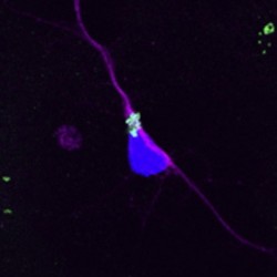
Research carried out by Nicole Gennet of the CSC’s Stem Cell Neurogenesis group and her collaborators has demonstrated successful delivery of functional proteins into mouse and human neural stem cells (NSCs), using treated polystyrene microspheres. Other groups have shown that microspheres can be taken up by cells, but this is the first time that delivery has been demonstrated in NSCs, which have proved difficult to transfect by alternative methods.
By attaching fluorescent markers to the spheres, Gennet and her co-workers were able to follow both the levels of internalisation of spheres into NSCs, and the rates of subsequent uptake of spheres by propagating cells as they divided over a period of three days. Fluorescence measurements of mouse NSCs indicated extensive initial internalisation of all three sizes of microspheres investigated – 200nm, 500nm and 2µm (2000nm) – but, for the larger microspheres, fluorescence per cell decreased after the first day. For the two smaller sphere sizes, no such decrease in fluorescence was recorded, indicating that the rate of uptake of the microspheres by the propagating cells made up for the drop in fluorescence per cell as the cells divided.
Next, the team assessed delivery of a biologically-functional molecule into mouse NSCs. Microspheres conjugated to the enzyme beta-galactosidase, which processes larger sugar molecules into monosaccarides, were introduced as before and their activity was followed by fluorescence flow cytometry and microscopy. Cells showed near total beta-galactosidase activity after a three day period.
The study also demonstrated microsphere activity in human NSCs, which exhibited extensive internalisation of the three microsphere sizes. In addition, mircospheres were also introduced into human NSCs just as they were induced to differentiate. Fluorescence measurements of two Morphologically-distinct cells made 30 days later demonstrated that both had internalised the mircospheres, indicating that their presence within a cell does not affect differentiation.
This work was published in New Biotechnology
Reference
Gennet, N, et al. Microspheres as a vehicle for biomolecule delivery to neural stem cells, New Biotechnology, (2009), in press.
The Edinburgh team that made the functionalised microspheres have published their work in Biomaterials:
Tsakiridis A, et al., Microsphere-based tracing and molecular delivery in embryonic stem cells, Biomaterials (2009), doi:10.1016/j.biomaterials.2009.06.024