Facility Staff
Academic Advisor
“The facility provides state of the art light microscopy equipment and image analysis tools as well as training and support for all LMS users.”
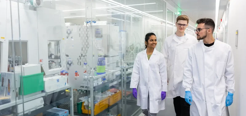
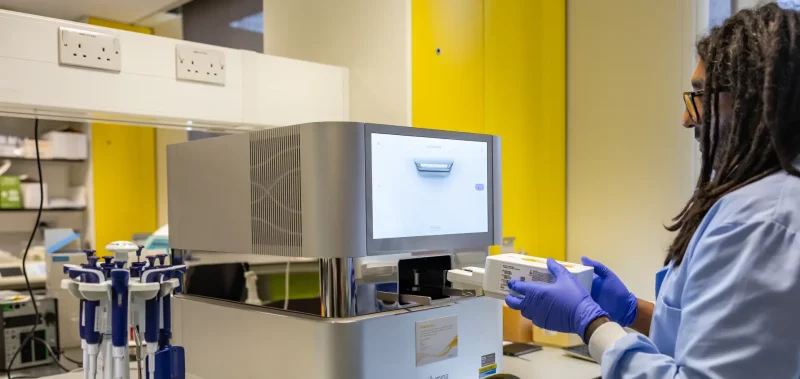
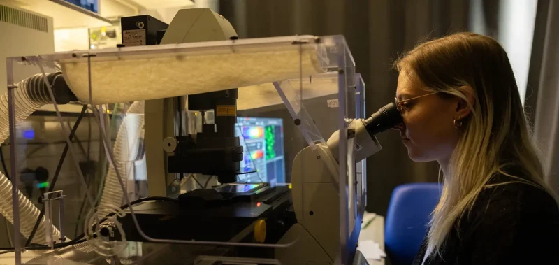
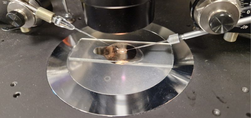
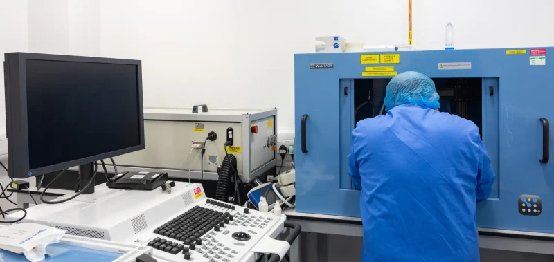
The Microscopy Laboratory offers a large variety of systems ranging from basic stereo microscopes to the latest confocal workstations to assist LMS staff and students in their research.
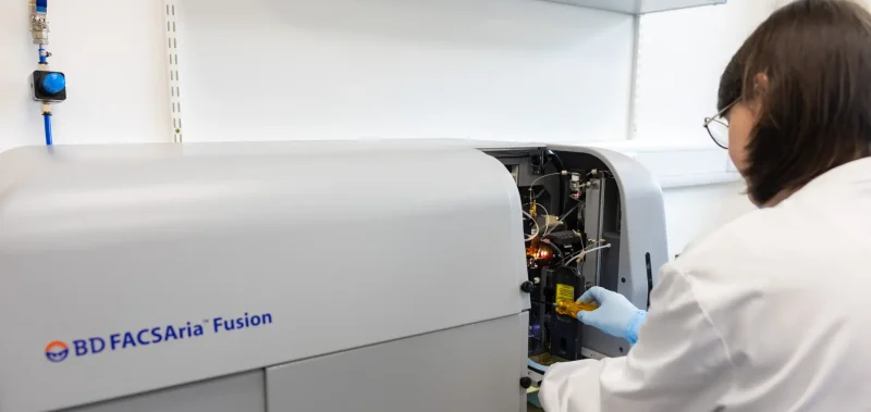
The facility is currently equipped with four confocal laser scanning microscopes, eight widefield fluorescence microscopes as well as super-resolution and light sheet instruments. Whole slide imaging is carried out on an AxioScan slide scanner and a PALM laser microdissection system is available for contact free sample cutting and capture. Live-cell imaging can be performed on seven workstations enclosed by environmental chambers to maintain constant temperature and CO2 levels. Several offline workstations can be used for image processing, visualisation and analysis of user data.
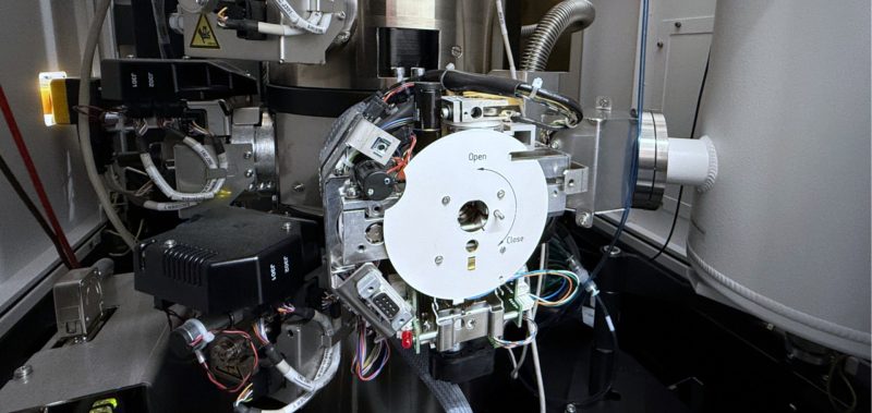
Staff provide user training and continuing support ranging from help with experimental design, image processing/visualisation and quantitative image analysis to direct collaboration on specific projects. An Electron Microscope (EM) and dedicated staff are also available to support projects examining cellular ultra-structure and single-particle structural analysis by cryo-electron microscopy (cryo-EM).
Find out more.
Figure 1. Liver section stained with DAPI and TRITC phalloidin
Figure 2. Two mouse fibroblast cell lines expressing H2B-EGFP or H2B-mCherry
Selected Publications
Sun, X. M., Bowman, A., Priestman, M., Bertaux, F., Martinez-Segura, A., Tang, W., Whilding, C., Dormann, D., Shahrezaei, V., Marguerat, S. (2020). Size-Dependent Increase in RNA Polymerase II Initiation Rates Mediates Gene Expression Scaling with Cell Size. Current Biology, 30, no. 7, 1217-1230.
Gutierrez-Escribano P, Newton MD, LlaurĂ³ A, Huber J, Tanasie L, Davy J, Aly I, Aramayo R, Montoya A, Kramer H, Stigler J, Rueda DS, Aragon L. (2019). A conserved ATP- and Scc2/4-dependent activity for Cohesin in tethering DNA molecules. Sci Adv 5, 6804.
Pollard, A. E., Martins, L., Muckett, P. J., Khadayate, S., Bornot, A., Clausen, M., Admyre, T., Bjursell, M., Fiadeiro, R., Wilson, L., Whilding, C., Kotiadis, V. N., Duchen, M. R., Sutton, D., Penfold, L., Sardini, A., Bohlooly, Y. M., Smith, D. M., Read, J. A., Snowden, M. A., Woods, A., Carling, D. (2019). Ampk Activation Protects against Diet Induced Obesity through Ucp1-Independent Thermogenesis in Subcutaneous White Adipose Tissue. Nature Metabolism, 1, no. 3, 340-349.
Dormann, D. (2019). Intensitycheck – the Light Measuring App for Microscope Performance Checks and Consistent Fluorescence Imaging. PLoS One, 14, no. 3: e0214659.