


By Lindsay Keith
November 8, 2021
Time to read: 3 minutes
To celebrate World Radiography Day 2021, we asked Ben Statton, MR Facility Manager at the MRC London Institute of Medical Sciences (MRC LMS) to tell us what it is like working as a radiographer in the LMS research environment.
The LMS MRI radiographers work alongside our researchers using the MRI facility. We are involved in every stage of the research process as an important member of the team.
As well as operating the MRI scanner itself, we are experts on a range of accessory equipment and software systems. This includes the functional MRI presentation system, patient physiological monitoring and pressure injectors, and the picture archive and storage system. By adhering to the study protocol we ensure the best possible images are acquired for every study. This is important for generating high quality, reproducible results.
Participants undergoing MRI are at the centre of everything we do, without them the research would not be possible. One of the most important aspects of our job is to ensure the participants and patients have a positive experience when they are involved in research.
After the participants have completed their MRI and left the department, the job of the radiographer is not finished. We must ensure all the acquired images are appropriately anonymised, and archived in a secure manner. In many cases the radiographers are also involved in the image post-processing, statistical analysis, and presentation of study results.
Currently, one of the research projects taking place at the Mansfield Centre for Innovation is using exercise cardiac MRI (CMR) – an innovative technique, undertaken at very few research centres internationally. It involves patients cycling on a pedal ergometer whilst undergoing an MRI scan of their heart.
Ben Statton, LMS MR Facility Manager, performing CMR at the Mansfield Centre for Innovation.
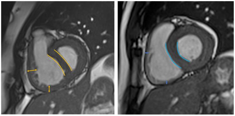
We are using exercise CMR to unmask right ventricular dysfunction in patients with pulmonary hypertension (PH). PH is a condition where pressure in the lungs is too high. As the condition progresses, the increase in lung pressure causes the right side of the heart to develop high pumping resistance. Eventually, this excessive cardiac resistance causes the heart to fatigue and fail.
In the early stages of PH, the right side of the heart can sometimes compensate. This enables the heart to pump normally when the person is at rest. However, during exercise, when the heart rate and workload are increased, the compensation may no longer be enough and this is when problems occur.
Currently, predicting outcomes for patients with PH is challenging, however imaging these patients while they are exercising may provide the additional information required to help predict survival.
At the LMS MR Facility we are using exercise CMR to identify new ways to measure and predict how a person’s heart responds to exercise. We are building upon research done in 2016 to translate this exercise CMR research method into clinical practice, with an aim for it to serve the PH service at Imperial College NHS Trust.