


A new study from the MRC Laboratory of Medical Sciences (LMS) reveals how ancient viral DNA once written off as “junk” plays a crucial role in the earliest moments of life. The research, published today in Science Advances, begins to untangle the role of an ancient viral DNA element called MERVL in mouse embryonic development and provides new insights into a human muscle wasting disease.
By Emily Armstrong
December 19, 2025
Time to read: 5 minutes
Transposable elements are stretches of DNA that can move around the genome. Many of these DNA sequences originate from long ago, when viruses inserted their genetic material into our ancestors’ genomes during infection. Today, these viral transposable elements make up around 8-10% of the mammalian genome.
Once disregarded as “junk” DNA, we now know that many transposable elements play an important role in influencing how genes are turned on and off, especially during early development. They have a variety of beneficial and harmful roles in the body, for example, some help regulate normal immune responses, while others can disrupt genes and contribute to diseases like cancer.
A central switch in early development
The latest research, led by Dr Michelle Percharde, Head of the Chromatin and Development group and former postdoctoral researcher Dr Paul Chammas, focuses on a viral transposable element called MERVL. This element becomes highly active for a short window of time when a mouse embryo reaches the 2-cell stage – the point at which a fertilised egg has divided into two cells and switches on its own genome for the first time. Cells in this state are considered ‘totipotent’, meaning they can generate every cell type of the embryo and extraembryonic tissues like the placenta.
MERVL acts as a central switch to activate a large network of genes specific to the 2-cell stage of development – but its precise contribution is not well understood.
Disentangling the network
To work out of the role of MERVL, the team used a gene manipulation technique called CRISPR activation to turn on MERVL elements in mouse embryonic stem cells, to mimic what happens in 2-cell embryos.
In cells where only MERVL was activated, the cells looked like they were only partially similar to cells of the 2-cell stage, but they still had several characteristics of totipotency. The researchers described this in-between state as an ‘intermediate phenotype’. They showed that activating MERVL alone is sufficient to create totipotent features in early embryonic development.
Transposable elements aren’t the bad guys
MERVL is activated by a transcription factor (a protein that helps genes turn on and off) called Dux. While Dux is important for switching on the 2-cell developmental programme, when it’s active for too long, it becomes toxic to cells and causes cell death.
To determine whether MERVL plays a role in these harmful effects, the team used the same CRISPR system to activate Dux, which they found activated MERVL but also lots of other genes. In this setting, they found that Dux activates a protein that triggers cell death, called Noxa. This confirmed that Dux, not MERVL, is responsible for some of the negative impacts on cells.
“By being able to compare what’s happening in these different contexts, we can see that transposable elements in this case aren’t the bad guys,” says Paul, “and we’ve successfully started to unpick some of the different roles different parts of the network play in early development.”
Implications for a muscle wasting disease
The human version of Dux is called DUX4. While it’s likely crucial for early development, it must be switched off permanently after this point. Certain genetic mutations cause the abnormal activation of DUX4 in adults which drives a disease called facioscapulohumeral muscular dystrophy (FSHD). In these patients, DUX4 causes problems such as cell death in the muscles, leading to progressive weakness and wasting.
When the team studied DUX4 in human cells, they found that DUX4 also increases levels of human NOXA. Interestingly, when they studied patient samples, they saw that those with the most severe forms of the disease had the highest levels of NOXA.
This suggests that NOXA could be a potential therapeutic target for this disease. Developing a drug to inhibit NOXA could prevent cell death, helping to improve survival of muscle cells in patients.
“More broadly, facioscapulohumeral muscular dystrophy is a complex disease – even though all cells of a patient have the genetic changes that cause it, only a subset of cells activate DUX4,” says Michelle, “Understanding what triggers DUX4 activation just in muscle cells, as well as how this compares to activation in early development, are key questions we hope to explore in future research.”
This study was funded by UK Research and Innovation (UKRI) and the Medical Research Council (MRC).
Read the full publication: https://www.science.org/doi/10.1126/sciadv.adu9092
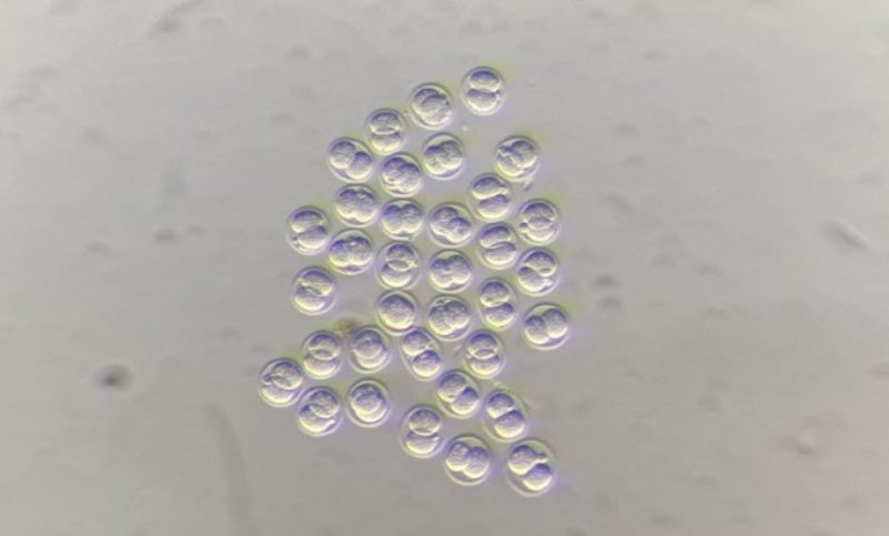
About the image at top of the page: A 2-cell stage mouse embryo visualised by fluorescence microscopy. Fluorescent molecules reveal different components within the cells. DNA is stained white, which reveals the nucleus of each cell. MERVL Gag protein (which is expressed because the embryo is at the 2-cell totipotent stage) is stained in blue and occupies the cytoplasm of each cell. Credit: Bryony Leeke, Postdoctoral Researcher, Chromatin and Development group.