By Helen Figueira
August 5, 2013
Time to read: 3 minutes
 Unlocking the structure of DNA helicase
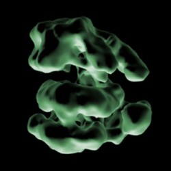
One of the key requisites for an organism to be considered living is its ability to multiply. Prior to cell division the cell’s DNA must be duplicated. At the molecular level this involves an incredibly well-regulated process common to all creatures from bacteria to humans. Without DNA replication, life as we know it wouldn’t exist. The procedure relies on two enzyme complexes: a DNA helicase splits the strands of DNA apart enabling the second enzyme complex, a DNA polymerase, to attach organic bases to the new strands. New research from the CSC’s DNA Replication Group reveals the structure of the helicase in complex with its DNA loading factors.
Group leader, Christian Speck, in collaboration with Huilin Li of Stony Brook University, New York, USA is using Cryo-electron microscopy (EM) to image the enzymes. This technique uses exceptionally low temperatures (-210c) to stabilise the molecules enabling incredibly high-resolution pictures to be taken. “It has a distinct advantage over other techniques, since it conserves the natural structure and folding of the proteins. High contrast is generated by Cryo-EM when the protein casts shadows onto itself,” explains Speck.
The researchers combined thousands of images together to generate a 3D representation of the complex. “However,” Speck reveals, “EM only gives you the outer shape.” To localise specific components of the large complex the team used fusion proteins, attached to clefts inside the helicase complex. “When the full picture of the protein came together we got our first ideas about its function. Without this structural model it’s like working in the dark.”
“We observed how the helicase and its loader interact,” explains Speck. “This gave us a clue about the interaction surfaces involved.” With this information the team can now explore the role of these interaction surfaces in helicase recruitment. “The function of the helicase loader is to open up the helicase ring,” confirms Speck, “then the DNA can slip inside.” He is excited to have gotten the first glimpse of this reaction, since it allows his team to watch DNA entering into the complex.
Uncovering the structural detail of this DNA replication complex has implications for drug design. Speck believes that DNA helicase inhibitors are potentially anti-cancerous. “We know that a natural inhibitor of helicase loading specifically kills cancer cells,” Speck reveals, “while causing cell cycle arrest in normal cells.” Currently no drugs of this type are on the market, but knowing the structure of the helicase’s intermediate state will undoubtedly drive investigation into potential therapies.
Knowing how DNA replicates is a fundamental biological question. This new picture of DNA helicase in complex with its loader is a big stepping-stone, and will fuel novel experimentation in the field. Could it be that we are finally unlocking the mechanism of what it takes to be considered “living”?
MT/BM
Reference
Jingchuan Sun,Cecile Evrin, Stefan A Samel, Alejandra Fernández-Cid,Alberto Riera, Hironori Kawakami, Bruce Stillman, Christian Speck & Huilin Li. Cryo-EM structure of a helicase loading intermediate containing ORC–Cdc6–Cdt1–MCM2-7 bound to DNA. Nature Structural & Molecular Biology 20,944–951(2013) doi:10.1038/nsmb.2629
Also featured in Nature’s article metrics