


By Helen Figueira
March 14, 2011
Time to read: 5 minutes
 How doing a twist helps DNA replicate
Twisted telephone handset wires are notoriously difficult to undo. Biology however uses this twisting or supercoiling effect to pack the genetic material tightly in the nuclei of cells. DNA is a right-handed double helix with one full turn being made for roughly every 10.5 base pairs. When the helix is twisted more or less tightly it causes the DNA to form supercoils, similar to twists in a telephone wire. If the DNA molecule is wound more tightly in the direction of the DNA helix (right handed) then the molecule is said to be positively supercoiled, whereas if the DNA is made less tightly wound (by applying a left-handed twist) it is said to be negatively supercoiled.
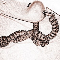
Positive and negative supercoiling causes the DNA to form loops or figure 8s, but in opposite directions (see graphic below). Many biological processes involve the opening of the DNA helix by specialised machines that rotate DNA – helicases. By their nature, helicases cause DNA supercoiling, which can cause problems for other cellular processes such as DNA replication. To resolve these problems cells employ specialised machines called topoisomerases, able to resolve the conformational alterations by breaking and rejoining DNA molecules.
In addition to changes to conformation occurring in the same DNA molecule, cells need to remove links that occur between different molecules of DNA, for instance between sister chromatids during DNA replication. A specialised topoisomerase (topoisomerase II) does this job by breaking one DNA molecule and passing the second DNA molecule through the break before rejoining the broken ends.
How doing a twist helps DNA replicate
Twisted telephone handset wires are notoriously difficult to undo. Biology however uses this twisting or supercoiling effect to pack the genetic material tightly in the nuclei of cells. DNA is a right-handed double helix with one full turn being made for roughly every 10.5 base pairs. When the helix is twisted more or less tightly it causes the DNA to form supercoils, similar to twists in a telephone wire. If the DNA molecule is wound more tightly in the direction of the DNA helix (right handed) then the molecule is said to be positively supercoiled, whereas if the DNA is made less tightly wound (by applying a left-handed twist) it is said to be negatively supercoiled.
Positive and negative supercoiling causes the DNA to form loops or figure 8s, but in opposite directions (see graphic below). Many biological processes involve the opening of the DNA helix by specialised machines that rotate DNA – helicases. By their nature, helicases cause DNA supercoiling, which can cause problems for other cellular processes such as DNA replication. To resolve these problems cells employ specialised machines called topoisomerases, able to resolve the conformational alterations by breaking and rejoining DNA molecules.
In addition to changes to conformation occurring in the same DNA molecule, cells need to remove links that occur between different molecules of DNA, for instance between sister chromatids during DNA replication. A specialised topoisomerase (topoisomerase II) does this job by breaking one DNA molecule and passing the second DNA molecule through the break before rejoining the broken ends.
“It has been long known that DNA replication causes the resulting sister chromatids to be linked or intertwined, explains Luis Aragon of the CSC Cell Cycle Group, “but for cell division to proceed, these intertwines need to be resolved, because sister chromatids must be physically separated. Although we know that topoisomerase II removes the links between sister chromatids, we also know that it is equally likely to introduce them. How its activity is regulated to ensure that its activity has a bias towards removal rather than introduction of links is one of the long-standing questions of mitosis.”
Jonathan Baxter and Nicholas Sen, researchers in Luis’s team, and their collaborators have discovered why topoisomerase II specifically removes links between sister chromatids during mitosis. The results of their study have recently been published in the journal Science.
Studying circular plasmids in yeast the scientists showed that when cells enter mitosis, DNA plasmids change their global topology and become highly wound or positively supercoiled. They then showed that in the mitotic topological state “topoisomerase II specifically removes links between sister DNA plasmid molecules, rather than introducing them or acting on individual plasmids to change their supercoiling status,” says Luis.
The team also found that cells have dedicated machines to promote the topological change associated with mitosis, called the Condensin complex. They propose a new model explaining how cells manage to ensure that topoisomerase II removes every single link between sister chromatids before chromosome segregation. These findings are medically relevant because many chemotherapeutic cancer treatments use inhibitors of topoisomerase II.
This research appears in Science
Reference:
Baxter, J., Sen, N., Martínez, V. L., De Carandini, M. E. M., Schvartzman, J. B., Diffley, J. F. X., Aragón, L., Mar. 2011. Positive supercoiling of mitotic DNA drives decatenation by topoisomerase II in eukaryotes. Science 331 (6022), 1328-1332.