


By Helen Figueira
April 22, 2016
Time to read: 7 minutes
Deborah Oakley
A group of sixth form science students visited the MRC Clinical Sciences Centre (CSC) in February to take part in the institute’s microscope workshops for local schools. It was the first time that A level students have taken part in the project.
The students had a unique opportunity to gain hands-on experience of using the high-resolution microscopes that they had been studying. With the help of CSC scientists, they had a close-up look at brain tissue, cancer cells and cells dividing in real time. For many of the students this was their first chance to see these tissues in real life, beyond the pages of their textbooks.
“I think the best bit was to actually use the microscopes, and the fact that it was related to the students’ syllabus. Any hands on experience is greatly appreciated,” said Cecile Roquain, the students’ biology teacher at St Charles College. “Having scientists talk about their work and how they use the equipment on a daily basis was great. Maybe we can arrange to come back soon!”
The visit began with a talk by Enrique “Fadri” Martinez-Perez, who leads the CSC’s Meiosis group. He explained how he uses microscopes in his research to explore the way in which our bodies’ cells make copies of the structures, called chromosomes, in which they store DNA. This copying process is called mitosis if it has one stage, or meiosis if it has two. The students had been studying both processes in school, and were interested to hear how Martinez-Perez is developing new techniques to study this in greater detail.
After the talk, they were able to see these techniques in action. Nuria Ferrandiz-Diaz, a postdoc in Martinez-Perez’s group, showed the students worms wriggling in a laboratory dish. She then introduced them to the powerful Delta Vision microscope, which she uses to observe the different stages of meiosis in worm cells. The students could focus in on individual cells and the squiggly chromosomes inside each of them. The way in which these chromosomes are lined up indicates which stage of meiosis a cell is in at any given moment. Ferrandiz-Diaz helped the students to identify the state of each cell.
David Tattersall, an Assistant Principal at St Charles College, said: “Cecile and the students really enjoyed it – looking at mitosis was particularly relevant to their studies.”
The students also got to grips with confocal and super-resolution confocal microscopes. For example, Dirk Dorman, head of the CSC’s Microscopy Facility, showed them how to use a microscope to zoom in on a particular protein inside a cell.
A team of young CSC researchers took the students on a tour of the different microscopes in small groups of three or four. This gave the students an opportunity to chat freely, in the absence of teachers. They talked about studying science at university and what life is like as a scientist. Meeting ‘real’ scientists in this way helped the students to appreciate that science is not restricted to those of a particular gender, background or ethnicity.
At the end of the workshop the students asked thoughtful questions about the possible results and applications of the research they had seen. Some were particularly interested to learn about Biomedical Picture of the Day (BPoD), one of the CSC’s public engagement initiatives. BPoD publishes an intriguing, strange or beautiful biomedical picture every day. Many of the students explored the BPoD website on their phones and began to follow it through their own social media accounts.
Younger generation
Also in February, around 90 children from local primary schools visited the CSC to use desktop light microscopes in what is becoming a traditional workshop. The Year 6 students extracted DNA from fruit and learnt about the genetics of eye colour.
A team of CSC volunteers welcomed one class of Year 4 children from Ark Conway Primary and two classes of Year 6 children from Old Oak Primary. Gareth Johnston, a teacher at Ark Conway Primary, said: “It’s fantastic to learn in a ‘real world’ environment. It will enable them to see the world in a new way, and to understand the connections between living things.”
The workshops began with a talk by Charlotte Mykura, in the CSC’s Cell Cycle group. Mykura explained that our bodies are made up of tiny building blocks, called cells, which stick together like Lego. She said that inside each cell is a set of instructions, called DNA. When she asked the children if they had heard about DNA and chromosomes, many shot their hands up to offer suggestions. She was surprised to see that even some of the Year 4 students had a general grasp of cellular biology.
The children then split into small groups to explore samples such as pondweed, blood and skin cells under the microscopes. CSC scientists were on hand to help, and the children drew diagrams of what they could see.
The station with fruit flies proved particularly popular. The children saw flies at different stages of their life cycle, including female flies whose bodies appeared to be bursting with eggs. “I liked looking at the fly transformation – it turns into little worms then flies,” said one of the Year 4 children.
Scientists from the CSC’s Gut Signalling and Metabolism group discussed how they use flies in their research. They explained that flies with particular characteristics, such as red eyes or curly wings, are easy to identify and this makes them easy to monitor in experiments. One of the Year 4 children said, “I liked looking at all the fruit flies because they are not all the same. Some had a lighter tone and some had a darker tone.”
The children were also thrilled to find out that a spoonful of yoghurt is teeming with life. One young girl said, “I liked the bacteria in the yoghurt. It was green and had lots of spots.” Another surprise came when they saw cancer cells for the first time. “I never thought it would look like that. I thought it would be the same as normal but just red,” said one student.
Some of the children brought their own samples of flowers, leaves and feathers to explore. One brought in some of his sister’s hair.
“The great staff team were very engaging and kept the children interested. Being in a real lab has been great, and using the equipment and talking to scientists has been even better. This was a fantastic trip!” said Chlorise Benjamin, a teaching helper from Ark Conway Primary.
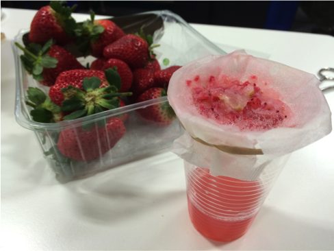
The Year 6 students also mushed up strawberries to extract the fruit’s DNA. This proved a messy but popular experiment that helped the students to learn more about how our bodies store genetic information. They particularly enjoyed taking home samples of the DNA sealed inside small test tubes.
They also wore colour-coded bracelets that they had made when learning about the different versions, or alleles, of genes. The bracelets were made using beads of four colours, one for each of the four possible letters, or bases, that make up DNA. Each student made a bracelet that coded for the combination of alleles that determine their eye colour.
For further information, contact:
Deborah Oakley
Science Communications Officer
MRC Clinical Sciences Centre
Du Cane Road
London W12 0NN
T: 0208 383 3791
M: 07711 016942
E: