By Deborah Oakley
A series of simple but important steps in bone microscopy has dramatically improved scientists’ ability to observe their inner structure, and the blood vessels that keep them healthy.
Pioneered by Saravana Ramasamy, who joined the MRC Clinical Sciences Centre’s (CSC) in August, this novel microscopy strategy could help researchers to better understand how our bones change as we age.
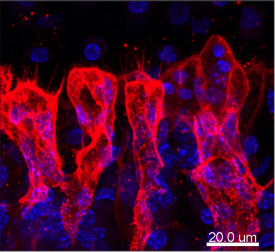
Blood vessels (red) are shaped like bulges at the end of a mouse’s tibia leg bone. The control centres, or nuclei, of the bone cells are shown in blue.
The first step is to chemically preserve bone tissue to keep its shape and structure intact. It’s then frozen and cut into thick slices. This allows the team to better visualise the bone’s 3D structure than has proved possible using the thin, flat slices of previous methods. Ramasamy has already spotted loops and bulges in the structure of the blood vessels that run through our bones, and this could be important in understanding how these vessels change with age.
He has used his new approach as part of a study to strengthen the bones of elderly mice. The findings, published in Nature in 2014, could help scientists to develop new treatments for elderly people whose fragile bones can break easily.
Speaking at his inaugural seminar on 14 September 2016, Ramasamy said it’s well known that as we age our bones lose mass and the ability to heal. “Around half of adults in Europe report musculoskeletal conditions. This is a big burden on the Government and the NHS, and it will only increase in the coming years, because of our sedentary lifestyles and ageing population.”
Ramasamy first began work on his imaging technique in 2014, whilst at the Max Planck Institute for Molecular Biomedicine in Germany. He joins the CSC to lead the new Integrative Skeletal Physiology group, and brings with him a hat-trick of grant successes.
The Wellcome Trust has awarded Ramasamy a Sir Henry Dale Fellowship, which helps young researchers to become independent scientists with their own groups. In August, he also received a Young Investigator Award from the American Society for Bone and Mineral Research to fund his presentation at a conference in Idaho, USA. Back in London, he’s been awarded a Junior Research Fellowship from Imperial College London. According to the College, this prestigious fellowship helps to “sustain the brightest and best early career researchers from across the world”.
Ramasamy will use these grants to explore how the blood vessels in our bones change with age. Blood vessels and bone tissue are intimately connected. Vessels in young bones “communicate” with the bone tissue and instruct it to grow. In return, the growing tissue prompts vessel growth. This positive feedback keeps the bones strong and healthy, but can be disrupted in the bones of older adults. According to Ramasamy, little has been known about this blood vessel-to-bone communication, and its disruption, because there has been no easy way to image the structure and inner layers of bone.
His microscope technique allows him to take a closer look. It can even zoom in on individual blood cells as they flow through living bone tissue in real time. It involves freezing and cutting thick slices of bone. The slices are then stained using fluorescent markers to easily identify blood vessels and bone cells (pictured above). Ramasamy published the details of his method last year and says it’s already being used by other researchers around the world.
He has used the approach to identify a previously unknown network of blood vessels in bone, known as ‘Type H’ vessels, because they have high levels of two particular proteins on their surface. Type H vessels are commonly found in young, growing bones and stimulate the bone to keep growing and build up mass. They do this by providing signals and molecules that are essential for cells that form bone tissue. According to Ramasamy, older bones have fewer Type H vessels, and this explains why bone mass decreases with age.
In a study at the Max Planck Institute, Ramasamy and colleagues showed that it’s possible to reactivate Type H vessels in aged bones by turning on a cellular signal called HIF. This reactivation promoted bone formation, which in turn restored bone mass in elderly mice. It could one day help scientists to develop a treatment for people. Ramasamy says the end goal of his research is to find new ways to treat bone and blood diseases that develop as we age.
To find out more get in touch:
Deborah Oakley
Science Communications Officer
MRC Clinical Sciences Centre
T: 0208 383 3791
M: 07711 016942
E: